
Surgery
Height Lengthening Surgery
Limb lengthening surgery was developed to help with leg straightening for children who were afflicted with growth abnormalities such as dwarfism and other deformities. Now it has become a cosmetic surgery for adults who have tired of being below average in height. Surgery to become taller can be a life-changing experience. It can increase your height from three to six inches. This procedure uses your body’s own miraculous healing process to increase your height. During the procedure, a surgical cut is made in one of the leg bones. An internal device is placed that will gradually spread the bone apart. The amazing part of this process is that your body will replace the empty space with tissue, connective tissue and blood vessels. Over time, the tissue hardens into bone and you will gain three to six inches in height.
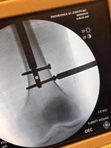
The first step in limb lengthening is to make a small incision in the front of the knee or by the hip and then surgically cut through the width of the bone to be lengthened (femur or tibia). Once the bone has been cut into two sections, the Stryde/Precice nail is fitted inside the center of the bone and secured with locking screws. The incision is then closed.
The next phase of this procedure can take several weeks to months to complete. It is called the distraction phase because the cut bone is gently pulled apart by a fraction of a millimeter at a time. Over time, the body creates new bone tissue to fill in the gap until the patient’s desired height is achieved. Nerves, blood vessels and tissue fill in the space between the bones and new bone begins to form and added height is achieved.
Since the leg bones contain bone marrow and stem cells, the best results occur when the bone is “seeded.” Seeding is accomplished by drilling several horizontal holes into the bone to expose the marrow. This allows the stem cells and red blood cells to saturate the surgical area to improve healing. Stem cells are specialized cells in the bone marrow that can perform many different healing functions. In limb lengthening, for example, stem cells can help form new bone, blood vessels, nerves, tendons, and ligaments.
As mentioned above, this “seeding” process also exposes bone marrow, which helps the body to produce new red blood cells. These RBCs help the recovery process by bringing oxygen-rich blood to the surgical area.
Height Lengthening specializes in limb lengthening surgery, a procedure for healthy adults who are unhappy with their height. We cater to individuals from all across the country seeking a permanent height increase solution to improve their stature, confidence, and overall quality of life. We utilize the PRECICE STRYDE™ Nail System to perform height enhancements for cosmetic purposes, which allows us to lengthen both legs simultaneously. We also specialize in bone lengthening surgery for those seeking bow leg correction or treatment for a knock knee deformity or leg length discrepancy.
Who is a candidate for cosmetic height surgery?
Candidates for cosmetic height surgery, also known as limb lengthening or stature lengthening, are typically healthy individuals who are dissatisfied with their current height and desire to increase it for aesthetic reasons. These individuals typically have reached skeletal maturity, usually around the age of 18 for females and 21 for males, and have realistic expectations regarding the outcomes of the procedure. Ideal candidates also include those who experience psychological distress or low self-esteem related to their height and want a permanent solution to enhance their appearance.
It is essential for height lengthening surgery candidates to undergo a thorough evaluation by a board-certified limb lengthening surgeon to ensure they meet the physical and psychological criteria for the procedure. Additionally, individuals should be willing to commit to the rigorous rehabilitation process and understand the potential risks and complications associated with the surgery. Overall, candidates for height lengthening surgery should be motivated to improve their self-confidence and quality of life through the enhancement of their stature.
While height increase surgery is life-changing for many individuals, it’s crucial for candidates to have a clear understanding of the risks, benefits, and potential outcomes associated with the procedure before proceeding.
How much taller can I get with bone lengthening surgery?
The amount of height increase limb lengthening surgery provides varies depending on several factors, including the patient’s initial height, bone structure, and treatment goals. Generally, height lengthening surgery provides height increases ranging from 2 to 6 inches or more.
Our height lengthening specialist recommends a total lengthening of 2-3 inches (5-8 cm) in the thigh bone (femur). Lengthening beyond 3 inches in a single bone increases the risk of complications, prioritizing patient safety. However, if desired, another limb lengthening surgery can be performed one year later in the shin bones (tibiae) to achieve an additional 2-3 inches (5-8 cm) of height.
It’s important to discuss your expectations and goals with a qualified surgeon during the consultation process. They can provide personalized recommendations based on your unique anatomy and desired outcome, helping you determine the potential height increase achievable through limb lengthening surgery.

How much does cosmetic height surgery cost?
The cost of cosmetic height surgery, also known as limb lengthening surgery, does vary significantly depending on various factors, including the specific procedure performed, the surgeon’s expertise, the facility’s location, and additional expenses such as pre-operative evaluations, post-operative care, and rehabilitation.
On average, the total cost of limb lengthening surgery can range from $80,000 to $100,000 or more per leg. At Height Lengthening, bilateral femur lengthening costs $82,000. Bilateral tibia lengthening costs $92,000, and combined tibia and femur lengthening costs $169,000. This cost typically includes the surgeon’s fees, anesthesia fees, hospital or surgical facility charges, and any associated medical expenses.
Another important cost consideration is rod removal. On average, the rod removal portion of height lengthening surgery costs $20,000-$25,000. Some insurances (primarily PPOs) help cover the cost of the rod removal bringing the cost down to $12,000-$15,000. To determine how much your health insurance contributes to the rod removal, verification must be performed one month before the procedure.
Also, Height Lengthening works with Care Credit. Patients who meet their credit criteria may qualify to finance their height lengthening surgery. Information for limb lengthening surgery financing is available during the consultation. We encourage individuals to explore all financing and health insurance options available to help make height lengthening surgery more affordable.
Is elective bone lengthening surgery covered by medical insurance?
It’s important to note that cosmetic height surgery is typically classified as an elective procedure and may not be covered by your health insurance. As a result, patients undergoing stature lengthening surgery are usually responsible for covering the full cost of treatment out of pocket. However, some insurers provide coverage for certain components of the procedure, such as the medications, physical therapy, and complications or additional issues that may arise from your limb lengthening surgery.
However, there are exceptions for certain circumstances. For example, if a patient has a medical condition that affects their height and causes functional impairment or psychological distress, insurance companies may consider covering the procedure as medically necessary. Additionally, if stature lengthening surgery is performed to correct a deformity or address a congenital condition that affects limb length, such as bow legs, insurance coverage may be more likely.
What happens during the initial consultation?
The height lengthening consultation is the first step of the height increase process. This appointment involves meeting with Dr. Mahboubian, to learn more about height lengthening surgery, how the procedure can help you achieve your height increase goals, and if the surgery is right for you. To determine if the procedure is right for you, we’ll carefully review your medical history, perform a physical exam. We’ll also take X-rays of your legs to calculate the dimensions necessary to fit the nail system properly to your bones. Dr. Mahboubian also discusses his treatment plan recommendation, addresses any expectations, and answers any questions you may have about your procedure.
Since many of our patients come from all over the country, we do offer online height lengthening surgery consultations for those unable to physically make it into our Burbank, California office. Keep in mind that there are limitations to the online consultation option, as it limits our ability to get the most accurate analysis of your situation.
*We reserve the online consultation option for individuals who cannot come to our office for an in-person consultation.
In-person limb lengthening surgery consultations are highly advised for optimal results. X-Rays are required for online consultations so we can order the right size nails for your treatment plan. The price of the in-person and online height lengthening surgery consultations are the same: $800.
With every one-on-one consultation, Dr. Mahboubian supplies his patients with a thorough understanding of their problem and a complete overview of their treatment options in order to allow his patients to make the most educated decisions about their care.
How do I schedule the surgery?
Scheduling information is available for individuals who are deemed candidates for height lengthening surgery.
Once a surgery date is confirmed, patients receive detailed instructions on pre-operative preparations, including any required medical tests, medications to avoid, and dietary guidelines. They’ll also receive guidelines on what to expect on the day of surgery, post-operative care instructions, and follow-up appointments.
Throughout the scheduling process, patients are encouraged to ask questions and communicate any concerns they may have to ensure a smooth and successful surgical experience.
Which bones will be lengthened?
Dr. Mahboubian performs both femur and tibia bone lengthening procedures. Due to the high rate of complication nerve damage, fat embolism, joint contracture, and other complications, lengthening is not done on both the femur and tibia bones simultaneously. However, patients interested in having both bone types elongated can have separate procedures scheduled several weeks apart.
View an overview of the limb lengthening process here.
How do you lengthen the bones and soft tissues?
Limb lengthening surgery starts with a small incision in the knee or hip, followed by surgically cutting the femur or tibia of the bone to be lengthened into two sections. Dr. Mahboubian inserts the Stryde/Precise nail into the center of the bone and secures it with locking screws before closing the incision.
The distraction phase occurs over the next several weeks to months. During this time, the surgically separated bone pieces are gradually stretched apart, a fraction of a millimeter at a time as the body generates new bone tissue , blood vessels, and nerves to fill the space created and add three to six inches to the patient’s height.
What is the PRECICE STRYDE™ Nail?
The PRECICE STRYDE Nail is a revolutionary new, less invasive internal limb lengthening device that can withstand up to 250 pounds of weight post-operatively. A key advantage of the PRECICE STRYDE Nail system is that it significantly reduces post-operative pain, discomfort, and patient recovery time.
The system is programmable. Prior to insertion, Dr. Maboubian pre-programs the device to gradually lengthen the patient’s bone to the prescribed length via remote controlled magnetic technology. A single PRECICE STRYDE Nail limb lengthening procedure provides up to a 3-inch height increase, while two procedures on each leg can yield a height increase of 6-inches.
What happens after the initial surgery?
After the initial limb lengthening surgery, patients undergo a period of post-operative recovery and rehabilitation. This typically involves a hospital stay of several days to monitor for any immediate complications and ensure proper wound healing. At this time, patients will also start physical therapy, which they will continue after discharge, during the rest of their height lengthening recovery.
Physical therapy plays a crucial role in the post-operative period, helping patients regain strength, mobility, and range of motion in the affected limb. Patients gradually increase their activity level and weight-bearing as tolerated, following a customized rehabilitation plan designed to optimize their recovery and ensure a successful outcome.
During the brief hospital stay, height lengthening patients will receive guidance on how to use the external remote control (ERC) to gradually lengthen the PRECICE rod in their leg(s) as directed by Dr. Mahboubian.
It’s important to note that height lengthening patients are not able to walk or bear weight on their legs during the first portion of the lengthening process and will require the use of mobility aids, such as wheelchairs, crutches, or scooters to get around. Regular follow-up appointments with Dr. Mahboubian, scheduled every 10 days to 2 weeks are scheduled to monitor progress via x-Rays and physical examination and assess healing, and make adjustments to the treatment as needed. Adjustments that may speed up or slow down the lengthening rate are determined on joint flexibility and bone healing progress.
Overall, the post-operative period is a critical phase of the limb lengthening process, requiring patience, dedication, and commitment to achieve optimal results.
Where do I stay during the lengthening process?
We recommend for limb lengthening patients to remain in Los Angeles for the duration of their recovery. However, we understand that may not be possible for some individuals. In that case, patients are encouraged to stay a minimum of two to three weeks to ensure no post-operative and lengthening complications. Anyone unable to remain in the area, should send us their x-Rays every few weeks or as advised by Dr. Maboubian while continuing their height lengthening recovery at home. We are available via phone, email, and chat, and can coordinate care with a local orthopedic surgeon near you.
What happens after I complete the lengthening process?
The complete height lengthening process takes up to 8 months to complete. This is the average amount of time it takes the bones to heal. During this time, patients will continue with physical therapy to strengthen their muscles to properly support their weight and regain their mobility. Most patients can safely resume their normal activities and undergo PRECICE Rod removal surgery within one year after insertion. The rod removal process is an outpatient procedure, so no hospital stay is required.
What are the potential limb lengthening complications?
However, limb lengthening is a surgical procedure that comes with unique, yet rare risks.
Infection: Surgical sites can become infected, leading to pain, swelling, redness, and fever. Prompt treatment with antibiotics is necessary to prevent complications.
Delayed bone healing (nonunion): Sometimes bones take longer than expected to heal, leading to delayed consolidation or non-union. Additional interventions, such as bone grafting, may be required to promote healing.
Nerve or blood vessel damage: During surgery, nerves and blood vessels near the bone may be injured, leading to numbness, tingling, impaired circulation, or deep vein thrombosis. Careful surgical technique and monitoring can help mitigate these risks.
Leg length discrepancy or Joint stiffness: Lengthening procedures can result in joint stiffness or contractures, limiting mobility and function. Physical therapy is essential to prevent and manage joint stiffness.
Hardware-related issues: Fixation devices such as external fixators or internal implants may cause discomfort, irritation, or complications such as loosening or breakage. Revision surgery may be necessary to address hardware-related issues.
Psychological distress: Lengthening procedures can be physically and emotionally demanding, leading to psychological distress such as anxiety or depression. Counseling and support services may be beneficial for patients undergoing limb lengthening surgery.
Dr. Mahboubian and the Height Lengthening care team’s orthopedic and limb lengthening expertise and unwavering commitment to delivering excellence in patient care ensure our patients have optimal outcomes.
What results have we seen after limb lengthening?
Many of our femur lengthening patients have added up to 3.1 inches (8cm) to their height with a single surgery. Those who also have limb lengthening surgery to their tibias gain an additional height increase of 2.5 inches (6.5 cm).
Why choose Height Lengthening for cosmetic height surgery?
Height Lengthening is a world renowned Los Angeles area based orthopedic and limb lengthening surgical center. Patients come from across the country to fulfill their height increase dreams and receive the best in orthopedic treatment by our compassionate care team. Our height increase surgeon and staff takes time to provide our patients with the care and support they need for a world-class height lengthening experience. All treatment plans are designed to meet the unique needs of our patients and help them achieve their height increase dreams.
Our Cosmetic Limb Lengthening Surgeon

Dr. Mahboubian was the pioneer orthopedic surgeon on the West Coast to perform cosmetic limb lengthening using the Precice and Stryde nail system. He practices the most cutting-edge techniques in orthopedic surgery, treating injuries in both adults and children, sports-related injuries, joint replacement surgery, hand injuries, neck and back pain, foot and ankle problems, and lower extremity deformities.
With a background in osteopathic medicine, Dr. Mahboubian has received specialized medical training focused on the musculoskeletal system, enabling him to provide comprehensive and meticulous care to his patients. Known for his friendly and professional demeanor, Dr. Mahboubian ensures that every consultation provides patients with a thorough understanding of their condition and treatment options, empowering them to make educated decisions about their care.
Driven by his passion for helping those with musculoskeletal injuries, Dr. Mahboubian discovered his fascination with limb lengthening and deformity correction towards the end of his orthopedic residency. Over the past few years, he has not only helped patients achieve their desired height and walk normally but has also boosted their confidence and enabled them to fulfill their lifelong goals.